Drawn to communicate science.

Clerkship E-Learning Module
E-Learning Module to Standarde Clinical Clerkship Training with Ovarian Neoplasms
The CHALLENGE
Clinical clerkships often vary in the level of student exposure to patients, depending on their geographical location. This inconsistency in hands-on experience can result in discrepancies in the accuracy of patient diagnoses and treatment plans. To address this, standardizing the training materials across clerkships was identified as a key solution. The goal was to improve student learning experiences and ensure that all students, regardless of their location, had access to the same quality of instructional content.
To test this solution, a pilot e-learning module was developed. The focus was on ovarian neoplasms—a topic critical for early diagnosis, yet challenging due to the limited exposure students may have to patients presenting with this condition. Notably, only 15% of ovarian neoplasm diagnoses occur at stage 1, underlining the importance of early detection. However, varied patient populations and a potential lack of exposure to typical presenting signs make this subject matter a challenge for students.
The Solution
I proposed the development of an interactive e-learning module to standardize and enhance the clerkship experience for students. The module was designed to improve the accuracy and efficiency of diagnoses by providing consistent, high-quality educational content across different clinical environments. The Ovarian Neoplasm Module was selected for the pilot due to its relevance and complexity in the context of early diagnosis.
Modules includes:
12 Neoplasm Models with Cross-Sections: Detailed anatomical models to demonstrate the structure and appearance of ovarian neoplasms from multiple perspectives.
1 Interactive 3D Pelvis Model: A dynamic 3D model of the pelvis, including the uterus and blood supply, to help students visualize the anatomical relationships involved in ovarian neoplasms.
1 3D Ovary and Follicle Model: A detailed 3D model to help students understand ovarian structure and the formation of neoplasms at the follicular level.
12 Illustrations: Depictions of ovarian neoplasms and related conditions, reinforcing key concepts.
7 Molecular Illustrations: Visual representations of the molecular mechanisms involved in ovarian neoplasms, helping students grasp the underlying of the condition.
10 Interactive Ultrasound Simulations: Interactive ultrasound images and simulations that allow students to practice identifying ovarian neoplasms in different clinical scenarios, further bridging the gap between theory and real-life application.
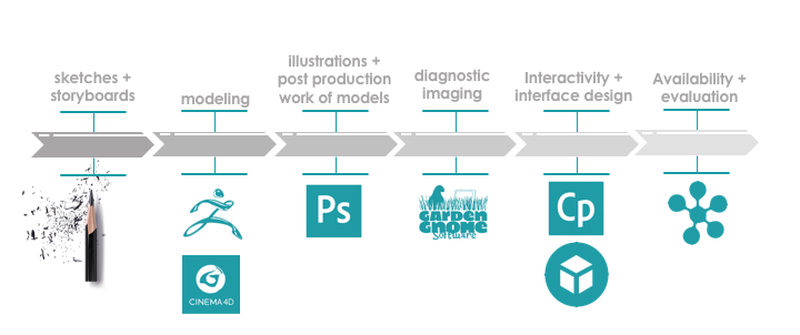
process
Needs Analysis & Collaboration with Subject Matter Experts (SMEs): I worked closely with clinicians, educators, and medical experts to identify the key learning objectives and critical concepts related to ovarian neoplasms. This collaboration ensured the module would cover essential content while addressing the diverse clinical environments in which students might train.
Content Development: I created detailed instructional content, including accurate 3D models, illustrations, and molecular diagrams, to visually support the material. These elements were designed to aid in understanding the complex pathophysiology and clinical presentation of ovarian neoplasms.
Interactive Learning Elements: To enhance student engagement and interactivity, I integrated several interactive features, such as 3D models, ultrasound simulations, and interactive quizzes. These elements were designed to promote active learning and ensure that students could apply their knowledge in realistic clinical scenarios.
Storyboarding & Visual Design: I developed a comprehensive storyboard to outline the flow of the module, ensuring that the multimedia components were well-organized and seamlessly integrated. This step helped to map out the user experience, providing a logical and effective progression of learning.
Pilot Testing & Feedback: A pilot version of the module was tested with a small group of students in different clerkship settings. Based on their feedback, I refined the module's content, interface, and interactivity to enhance the overall learning experience and ensure it met the students' needs.